Ivan Gerdzhikov
Department of Prosthetic Dentistry, Faculty of Dental Medicine, Medical University – Sofia, Bulgaria.
ABSTRACT:
Introduction: Operative treatment of tumours of the maxilla impairs the barrier between the oral and nasal cavi- ties, making difficult or impossible to eat, speak, and drink liquids.
Aim: The purpose of the clinical case described, is to examine the possibility of prosthetic treatment in pa- tients with fully resected hard palate, the treatment effect and recovery of impaired functions.
Materials and methods: The article presents the prosthetic treatment of a patient with resection of the en- tire hard palate as a result of an oncological disease. Due to complete edentulism, the treatment plan included the construction of an obturator and a complete lower denture. Functional impressions with additive silicone were taken with custom trays of the light-cured acrylic resin after pre- edging with wax impression material (ISO Functional GS). The dentures were made of a colourless heat cured the acrylic resin. The volume and localization of the defect ne- cessitated a specific shaping of the replacement part of the obturator. In order to provide the necessary retention and stability, we used our own modification of the open cup- shaped form in which the edges of the replacement part were extended distally in the area of the soft palate.
Results: The results of the treatment showed good retention and stability of the obturator and the lower den- ture. The feeding, speech and swallowing of the patient were successfully restored.
Conclusion: Prosthetic treatment with a definitive obturator allows optimal sealing of the maxillary defect even with a fully removed hard palate.
Keywords: maxillary resection, maxillary defect, obturator, post resection denture,
INTRODUCTION
Studies show a significant increase of oncological diseases in the maxillofacial area, with a tendency for the continuous increase [1, 2]. Some data suggest that oral can- cer is the most prevalent oncology disease after lymphoma and leukaemia [3]. Four-fold increased morbidity gives Suba et al. [4] grounds to define oral cancer as the disease of the 21st century.
Prosthetic treatment methods have a central place in the complex treatment and rehabilitation of patients with maxillary resection [5, 6]. Most authors [6, 7] conduct pros- thetic rehabilitation after maxillary resection in three stages, by means of a surgical, temporary and definitive obturator. Each of these prostheses allows the recovery of damaged functions through the various stages of treatment, ensur- ing the maintenance of a relatively good quality of life [8]. Prosthetic treatment of patients with maxillary resection is accompanied by many difficulties and problems associated with providing a stable barrier between the oral and nasal cavities and the sealing of the defect [9]. This necessitates preliminary planning of the prosthetic con- struction in accordance with both the basic prosthetic prin- ciples and the individual characteristics of the patient [10]. It is necessary to take into account all factors that influ- ence the retention, and stability of the obturator [11].
There are a number of methods for making defini- tive obturators with different materials and technologies [12, 13, 14]. Some authors [15] give priority to treatment with closed hollow obturators. According to others [16], treat- ment with hollow or dense buccal flange obturators is as- sociated with a number of difficulties, requiring their re- placement by open obturators. Their main advantage is the reduced weight – from 6.55% to 35.06% less than dense obturators [17]. Electromyographic studies show better clinical results when using the open cup-shaped form of the replacement part [18]. This is explained by reduced weight and volume, making it easier to put in the defect, and providing greater comfort for patients [16]. The main disadvantage of open cup-shaped obturators is plaque con- tainment and difficult cleaning [19].
AIM
The purpose of the clinical case described is to ex- amine the possibility of prosthetic treatment in patients with a fully resected hard palate, and the effect of treat- ment for the recovery of impaired functions.
MATERIALS AND METHODS
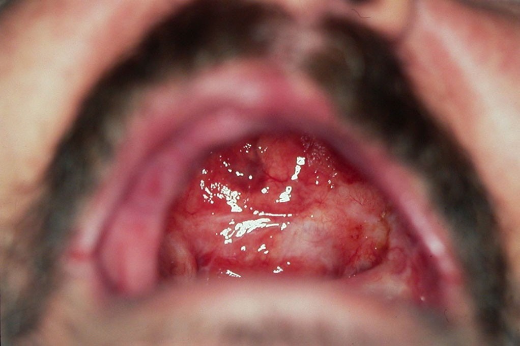
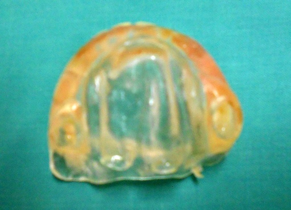
The article describes the prosthetic treatment of a 58-year old patient with resection of the maxilla as a re- sult of surgical treatment of oncological disease. Intraoral examination showed a large maxillary defect involving the whole hard palate and passing the A-line [Fig. 1]. The bro- ken barrier between the mouth and the nasal cavity did not allow normal feeding and speaking of the patient. It was impossible to drink liquids, which, according to the patient, was a major problem that had a serious impact on his qual- ity of life. The atrophy of the preserved alveolar ridge and the lack of natural teeth made it difficult to plan and carry out the prosthetic treatment. A treatment plan was com- piled, which included the construction of an obturator and a complete lower denture. The preliminary impression of the maxilla was taken with a standard metal tray elongated in the area of the soft palate defect. We used an irrevers- ible hydrocolloid impression material after preliminary tam- ponage of the defect with a gauze. Individual trays of light- cured acrylic were used for taking functional impressions of the two jaws. For the upper jaw, the tray was further shaped so that it’s edges entered circularly about 5 mm in the defect. This allowed preliminary edging both on the valve area and on the defect boundary for which ISO Func- tional (GS) wax was used. An additive silicone was used for finishing off the functional impressions. The occlusion height and the central position of the lower jaw were fixed with wax rims. After a successful trial denture, the dentures were finished in a colourless heat-cured acrylic resin. The volume and localization of the defect necessitated a spe- cific shaping of the replacement part of the obturator. To ensure the necessary retention and stability, we used our own modification of the open cup-shaped form in which the edges of the replacement part were extended distally in the area of the soft palate [Fig. 2]. This created a pre- requisite for additional sealing in the A-line area and sta- bilized the obturator during eating and swallowing. The dentures were adjusted and articulated in the last clinical stage. At the follow-up examinations, the occurred decu- bital injuries were treated, and some occlusal contacts were corrected.


RESULTS
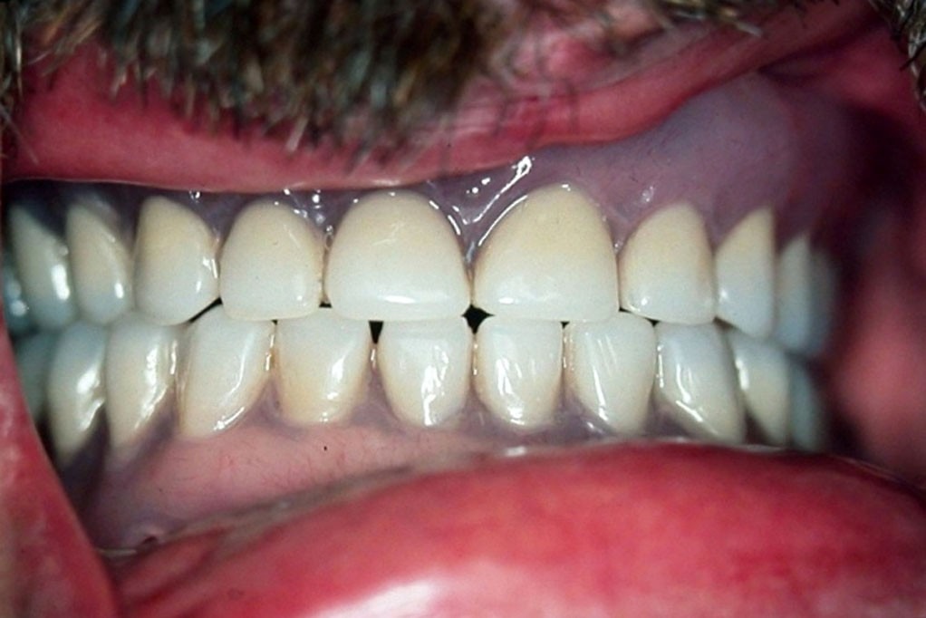
The results of the treatment showed good retention and stability of the obturator and the lower denture despite the large size and unfavourable localization of the defect. The modification used to make an open obturator with distal wings to the soft palate allowed optimal sealing and ensures successful recovery of the feeding, speech and swal- lowing of the patient. The patient’s main problem related to the inability to take liquids was also solved. The com- plex prosthetic treatment with an obturator and a lower denture allowed restoration of the occlusal relationships and the achievement of bilaterally balanced occlusion [Fig. 3]. Recovery of impaired functions regained the patient’s self-esteem and significantly improved his quality of life.

DISCUSSION
The applied treatment method confirmed the con- cept that prosthetic treatment has a central place in the com- plex treatment and rehabilitation of patients with maxil- lary resection [5, 6]. Proper planning and compliance with the patient’s individual characteristics allowed to make an optimal prosthetic construction, which according to most authors [10, 11] is important for the success of the treat- ment. The use of the classical open form of the replacement part provided easy insertion into the defect and con- firmed the advantages of this type of obturators [16, 18]. Reduced weight has helped for easier and quicker adapta- tion to the prosthesis, as reported by other authors too [17]. These advantages have had a beneficial effect on the pa- tient’s social life and activity as well as on his overall qual- ity of life, which according to most authors [8, 9] is the main goal of prosthetic rehabilitation.
CONCLUSION
Prosthetic methods of treatment are a major means of restoring speech, feeding and breathing for patients with maxillary resection. The variety of defects requires the ap- plication of specific methodologies and modifications, the aim of which is to achieve optimal sealing and create a sta- ble barrier between the oral and nasal cavities.
REFERENCES:
- Lung T, Tascau O, Almasan H, Muresan O. Head and neck cancer, epi- demiology and histological aspects – Part 1: a decade’s results 1993-2002. J Craniomaxillofac Surg. 2007 Mar; 35(2):120-5. [PubMed]
- Flores-Ruiz R, Castellanos- Cosano L, Serrera-Figallo MA, Gutiérrez-Corrales A, Gonzalez-Martin M, Gutiérrez-Pérez JL, et al. Evolution of oral cancer treatment in an andalu- sian population sample: Rehabilitation with prosthetic obturation and remov- able partial prosthesis. J Clin Exp Dent. 2017 Aug;9(8):1008-14. [PubMed]
- Al-Balawi SA, Nwoku A. Manage- ment of oral cancer in a tertiary care hospital. Saudi Med J. 2002 Feb; 23(2):156-9. [PubMed]
- Suba Z, Mihályi S, Takács D, Gyulai-Gaál S. [Oral cancer: morbus Hungaricus in the 21st century.] [in Hungarian] Fogorv Sz. 2009 Apr; 102(2):63-8. [PubMed]
- Artopoulou II, Karademas EC, Papadogeorgakis N, Papathanasiou I, Polyzois G. Effects of sociodemogra- phic, treatment variables, and medical characteristics on quality of life of pa- tients with maxillectomy restored with obturator prostheses. J Prosthet Dent. 2017 Dec;118(6):783-789. [PubMed] [Crossref]
- Chen C, Ren WH, Huang RZ, Gao L, Hu ZP, Zhang LM, et al. Quality of Life in Patients After Maxillectomy and Placement of Prosthetic Obturator. Int J Prosthodont. 2016 Jul-Aug;29(4):363-8 [PubMed] [Crossref]
7. Huryn JM, Piro JD. The maxillary imediate surgical obturator prosthesis. J Prosthet Dent. 1989 Mar;61(3):343-[PubMed]
8. Ali MM, Khalifa N, Alhajj MN. Quality of life and problems associated with obturators of patients with maxillectomies. Head Face Med. 2018 Jan 5;14(1):2. [PubMed] [Crossref]
9. Mittal M, Sharma R, Kalra A, Sharma P. Form, Function, and Esthe- tics in Prosthetically Rehabilitated Maxillary Defects. J Craniofac Surg. 2018 Jan;29(1):e8-e12. [PubMed] [Crossref]
10. Keyf F. Obturator prostheses for hemimaxillectomy patients. J Oral Rehabil.2001 Sep;28(9):821-9. [PubMed]
11. Parr GR, Gardner L. The evolution of the obturator framework design. J Prosthet Dent. 2003 Jun;89(6):608- 10[PubMed] [Crossref]
12. Tasopoulos T, Kouveliotis G, Polyzois G, Karathanasi V. Fabrication of a 3D Printing Definitive Obturator Prosthesis: a Clinical Report. ActaStomatol Croat. 2017 Mar;51(1):53-58. [PubMed] [Crossref]
13. Mawani DP, Muddugangadhar BC, Das A, Kothari V. Flasking tech- nique with alum crystals for fabricating definitive hollow bulb obturators. J Prosthet Dent. 2018 Jul;120(1):144-146. [PubMed] [Crossref]
14. Mohamed K, Mani U, Saravana- kumar P, Kumar SP, Arunachalam R. Split Hollow Bulb Obturator to Reha- bilitate Maxillary Defect: A Case Re- port. Cureus. 2016 Jun 9;8(6):e635. [PubMed] [Crossref]
15. Worley JL, Kniejski M. A method for controlling the thickness of hollow obturator prostheses. J Prosthet Dent. 1983 Aug;50(2):227-9. [PubMed]
16. Oh WS, Roumanas E. Optimiza- tion of maxillary obturator thickness using a double-processing technique. J Prosthodont. 2008 Jan;17(1):60-3. [PubMed]
17. Wu YL, Schaaf N. Comparison of weight reduction in different designs of solid and hollow obturator prosthe- ses. J Prosthet Dent. 1989 Aug;62(2): 214-7. [PubMed]
18. Hasanreisoglu U, Gürbüz A, Beyazova M. [Electromyographic evaluation of different types of obtura- tors constructed after maxillary resec- tions.] [in Turkish] Ankara Univ Hekim Fak Derg. 1989 May;16(1):45-51. [PubMed]
19. Asher ES, Psillakis J, Piro J, Wright R. Technique for quick conver- sion of an obturator into a hollow bulb. J Prosthet Dent. 2001 Apr;85(4):419- 20 [PubMed] [Crossref]
Please cite this article as:Gerdzhikov I. Prosthetic treatment of patient with total hard palate resection. J of IMAB. 2019 Jan-Mar;25(1):2355-2357. DOI: https://doi.org/10.5272/jimab.2019251.2355
Address for correspondence:
Dr Ivan Gerdzhikov, Department of Prosthetic Dental Medicine, Faculty of Dental Medicine, Medical University – Sofia; 1, St. G. Sofiyski blvd., 1431 Sofia, Bulgaria e-mail: ivan_ger1971@abv.bg
